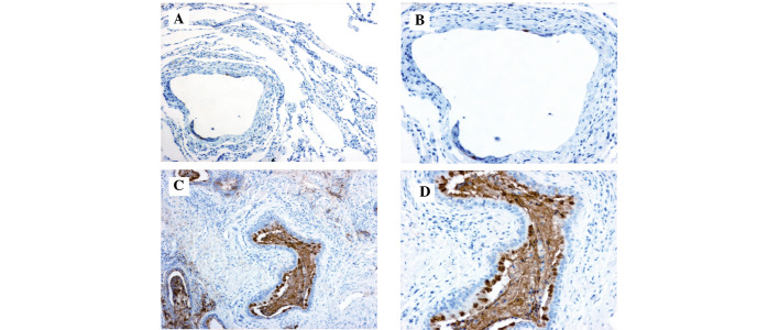
Figure 4.
Immunohistochemical staining demonstrated (A and B) weak staining of carbohydrate antigen 19-9 in the normal lung tissue [magnification, (A) ×100, (B) ×200] but (C and D) marked staining in the sequestrated lung tissue, particularly in the mucus of the cysts. [Magnification, (C) ×100, (D) ×200]. The reaction was amplified using the streptavidin-biotin-peroxidase method and diaminobenzidine was used as a chromogen.

