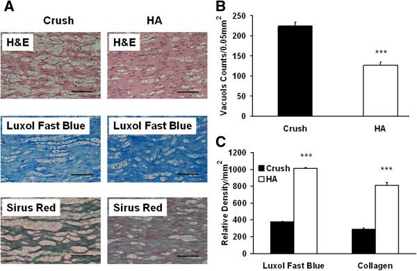
Figure 2.

Vacuole counts, Luxol fast blue, and Sirus red staining 4 weeks after hyaluronic acid treatment. After neurobehavioral assessment and electrophysiological examination, the injured nerve were harvested for assessments (A) Representative photomicrographs illustrating vacuole, Luxol fast blue, and Sirus red staining in each group, (B) Quantitative analyses of vacuole counts, (C) Quantitative analysis of, Luxol fast blue, and collagen density. ***p < 0.001; Bar length = 50 μm, n = 6.
