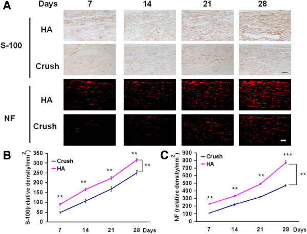
Figure 4.
Expression of nerve regeneration plotted against with time course. The injured nerves were harvested at various time points of 7, 14, 21, and 28 days after nerve crush. These nerves were subjected to immunohistochemistry staining of S-100 and neurofilament. (A) Microphotograph showed the expression of S-100 and NF in different treatment related to various time points. (B) Quantitative analysis of S-100 at different time points (C) Quantitative analysis of NF at different time points. **p < 0.01; ***p < 0.001, Bar length = 100 μm; n = 3. HA and NF: see text.

