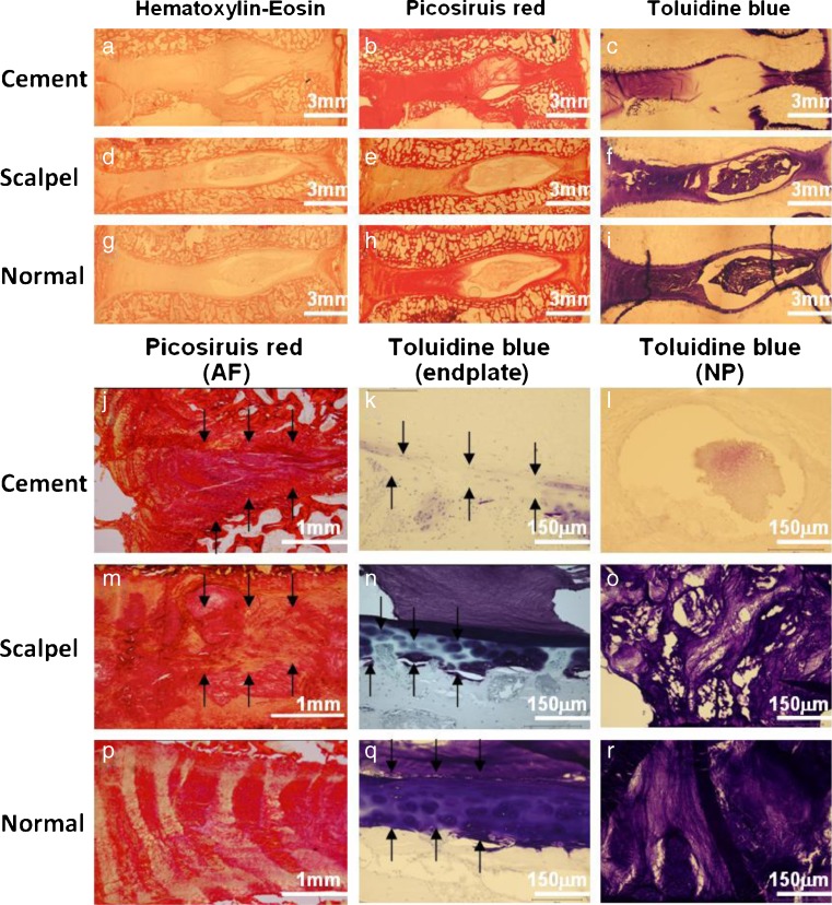
Fig. 5.
Histological images of discs in different interventions. (a-i) Overview of the discs in different histological staining. (j–r) Images with higher magnification. Picosiruis red staining viewed under polarized light: (j) irregular and collapse AF (arrow); (m) irregular fibrous lamellae (arrow); (p) regular fibrous lamellae. Toluidine blue staining: (k) disorganized endplate with loss matrix (arrow); (n) focal thinning of the EP (arrow); (q) uniformly thick EP with organized ECM; (l) very few matrix in NP (arrow); (o) matrix loss in NP; (r) normal matrix in NP

