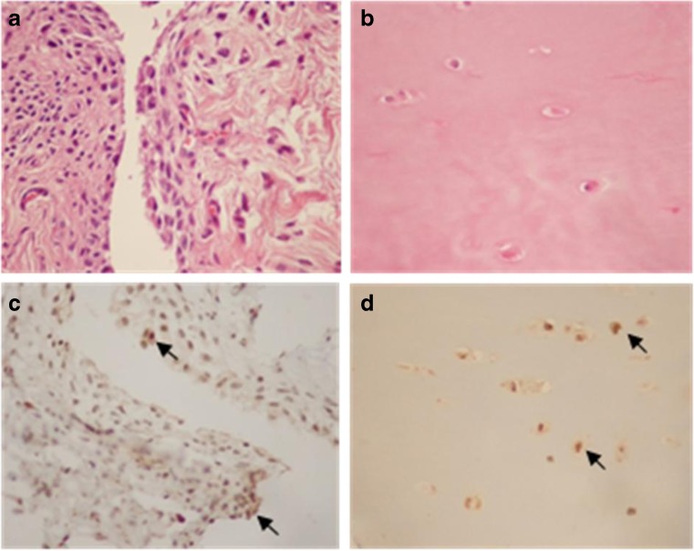
Fig. 4.
Representative photomicrographs of synovium and articular cartilage samples from knee OA patients. a, b Haematoxylin and eosin staining in synovium and articular cartilage, respectively. c, d VEGF immunohistochemical staining in synovium and articular cartilage of knee OA patients demonstrated the presence of VEGF expression in synovial lining cells and chondrocytes (arrows), respectively (×400)

