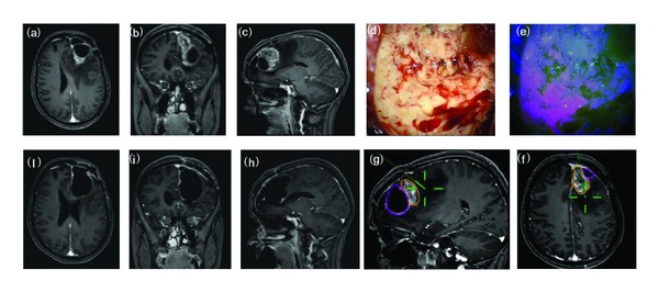
Figure 3.

F/43Y, preoperative MRI scan with gadolinium showing a recurrent frontal glioblastoma (a–c). Intraoperative view: tumor resection under white (d) and blue (e) light. Intraoperative boundaries of surgical cavity at MRI navigation after resection under blue light (f-g). Postoperative MRI scan with gadolinium (h–j) showing the complete tumor resection and the postsurgical cavity roughly larger than tumor size at preoperative imaging. Histological report: recurrent glioblastoma (astrocytoma grade IV sec WHO).
