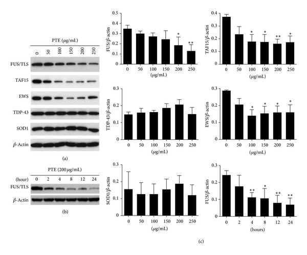
Figure 2.

Decreased FET family protein expression by PTE in SK-N-SH cells. (a) Cells were treated with different concentrations of PTE for 24 h; the expression of FUS/TLS, EWS, TAF15, TDP-43, SOD1, and β-actin was detected by Western blot. (b) Cells were treated with 200 μg/mL PTE for different time periods; Western blot was performed. (c) The Western blot results were quantified and statistical analysis was performed. Values are mean ± SEM of three independent experiments. *P < 0.05, **P < 0.01, and ***P < 0.001 compared with untreated control.
