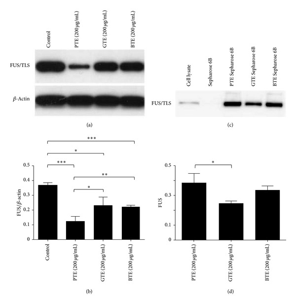
Figure 3.

Effects of three different tea extracts on FUS/TLS in SK-N-SH cells. (a) Cells were treated with 200 μg/mL PTE, GTE, or BTE for 24 h; the expression of FUS/TLS was detected by Western blot. (b) The Western blot results were quantified and statistical analysis was performed. Values are mean ± SEM of three independent experiments. *P < 0.05, **P < 0.01, and ***P < 0.001 compared with untreated control. (c) Cell lysates were incubated with PTE Sepharose 6B, GTE Sepharose 6B, BTE Sepharose 6B, or Sepharose 6B beads, and the levels of bound FUS/TLS were analyzed by Western blot. (d) The Western blot results were quantified and statistical analysis was performed. Values are mean ± SEM of three independent experiments. *P < 0.05.
