
Figure 6.
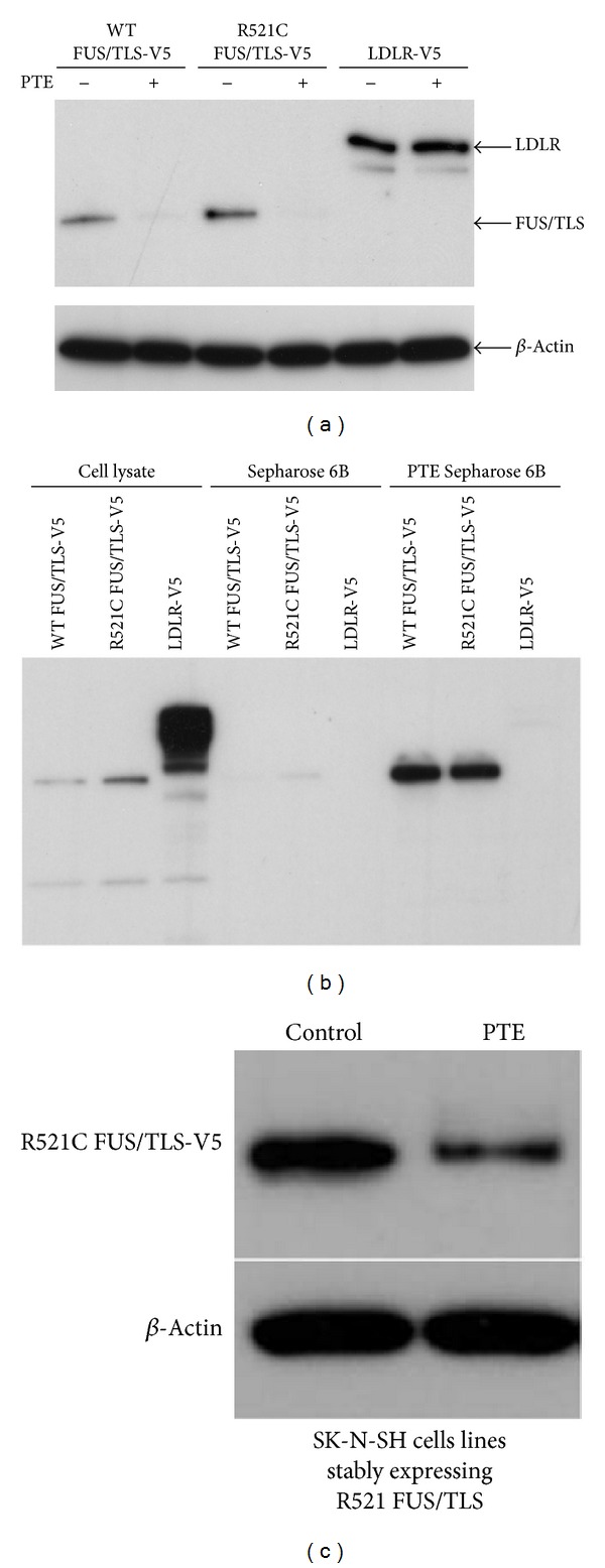
Degradation of R521C FUS/TLS protein by PTE. (a) SK-N-SH cells were transfected with V5-tagged wild-type FUS/TLS or R521C FUS/TLS or LDLR. Transfected cells were treated with or without PTE (200 μg/mL) for 24 h. Cell lysates were detected by Western blot with anti-V5 antibody. (b) HEK 293T cells were transfected with V5-tagged wild-type FUS/TLS, R521C FUS/TLS, or LDLR. Protein extracts from transfected 293T cells were incubated with PTE Sepharose 6B or Sepharose 6B beads. Proteins that were bound to the beads were analyzed by Western blot with anti-V5 antibody. (c) SK-N-SH cells stably expressing R521C FUS/TLS were treated with or without PTE (200 μg/mL) for 24 h. Cell lysates were detected by Western blot with anti-V5 antibody. β-actin was used as the loading control.