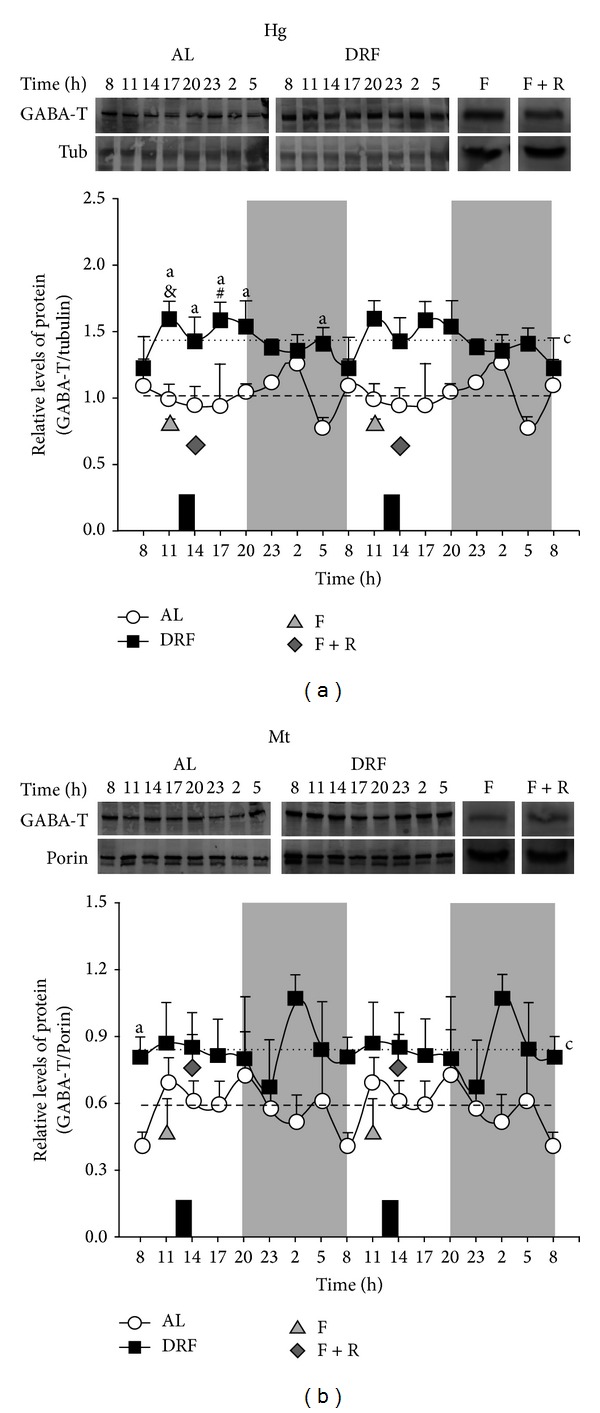
Figure 1.

Western blot analysis of 24 h profile of GABA-T protein in liver of AL and DRF rats. Liver homogenates (Hg) (a) and mitochondrial fractions (Mt) (b) were subjected to electrophoresis on 10% SDS-polyacrylamide gels. Data were quantified by densitometry of the bands obtained from homogenates (a) and mitochondrial fractions (b) collected over a 24 h period from the AL and DRF groups and food condition controls, F and F + R. Food availability for the DRF group is indicated by dark boxes (from 12:00 to 14:00 h). Average values are represented as a dashed line for the AL group and as a dotted line for the DRF group. Graphs represent the mean ± SEM of 4 rats per time point. Significant differences (P < 0.05) are indicated as follows: a: DRF versus AL by two-way ANOVA and post hoc Bonferroni test; c: mean of DRF versus mean of AL by Student's t-test for both homogenate and mitochondrial fraction; for homogenates only: &, DRF versus F at 11:00; #, DRF versus F + R at 14:00, both by the Student's t-test.
