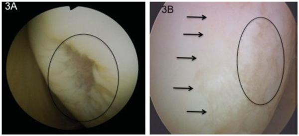
Figure 3.
Lesion 3A is an isolated Grade 3b defect in an otherwise pristine appearing knee joint. This patient went on to receive a microfracture and did well. Figure 3B is a similar size Grade 3b lesion (indicated with the circle) surrounded by areas of Grade 2 lesions. This patient failed an initial microfracture and went on to receive an autologous chondrocytes implantation involving the majority of her condyle (2.2×4.8cm). Even though this is obvious on the video of this lesion it is difficult to document this significant difference in character of this lesion in pictures.

