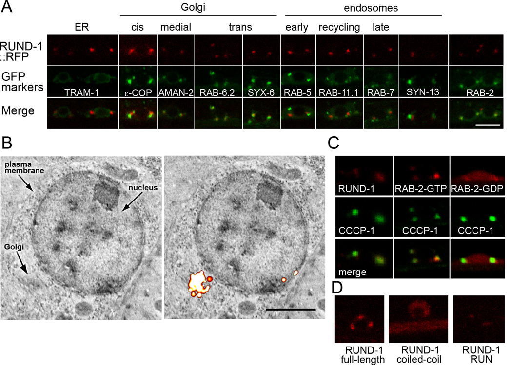
Figure 5. RUND-1 colocalizes with RAB-2 at the trans-Golgi network.
(A) RUND-1 colocalizes with RAB-2 and trans-Golgi markers. Each panel shows a single slice of a confocal image of motor neuron cell bodies in the ventral cord of young adult animals. The top boxes show the localization of a single-copy rescuing RUND-1::tagRFP-T fusion protein (oxIs590). RUND-1 localizes almost exclusively to two or three perinuclear puncta per cell. The middle boxes show the localization of single-copy GFP-tagged compartment markers. The bottom boxes show the merged images. Scale bar: 5 µm, applies to all panels. RUND-1 shows tightest colocalization with RAB-6.2, a trans-Golgi Rab protein, and with SYX-6, the ortholog of the trans-Golgi SNARE syntaxin 6. RUND-1 puncta also colocalize well with RAB-2 puncta. RUND-1 partially overlaps with the medial-Golgi marker mannosidase II (AMAN-2) and the cis-Golgi marker εCOP. RUND-1 is not colocalized with the rough ER marker TRAM-1 nor with the endosomal markers RAB-5, RAB-11.1, RAB-7 and SYN-13.
(B) RUND-1 localizes to the Golgi. The left panel shows a backscatter scanning electron micrograph of the cell body of a neuron in the nerve ring. The right panel shows the same image overlaid with the corresponding fluorescence PALM image of RUND-1::tdEos. Scale bar: 1 µm.
(C) CCCP-1 colocalizes with RUND-1 and RAB-2. CCCP-1 colocalizes with RUND-1 and RAB-2(GTP). CCCP-1 is still punctate when coexpressed with RAB-2(GDP), which is diffusely expressed.
(D) The RUN domain of RUND-1 mediates its localization. Full length RUND-1, the coiled-coil domain, and the RUN domain were tagged at their C-termini with tagRFP-T and integrated in the genome. The truncated proteins were expressed at lower levels. See also Figure S8.

