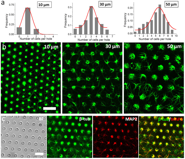
Figure 3. Culture of hippocampal neuron in NeuroArray.
(a) Distribution of neuron cells in NeuroArray with through-holes of different diameters. (b) Florescence images of βIII-tubulin showing the growth of neuron cells in NeuroArrays for 6 days. (c) Characterization of long-term neuron culture (18DIV) in NeuroArray of 30 µm through-holes. Cells were stained for βIII-tubulin (green) and MAP2 (red). Scale bar, 100 µm.

