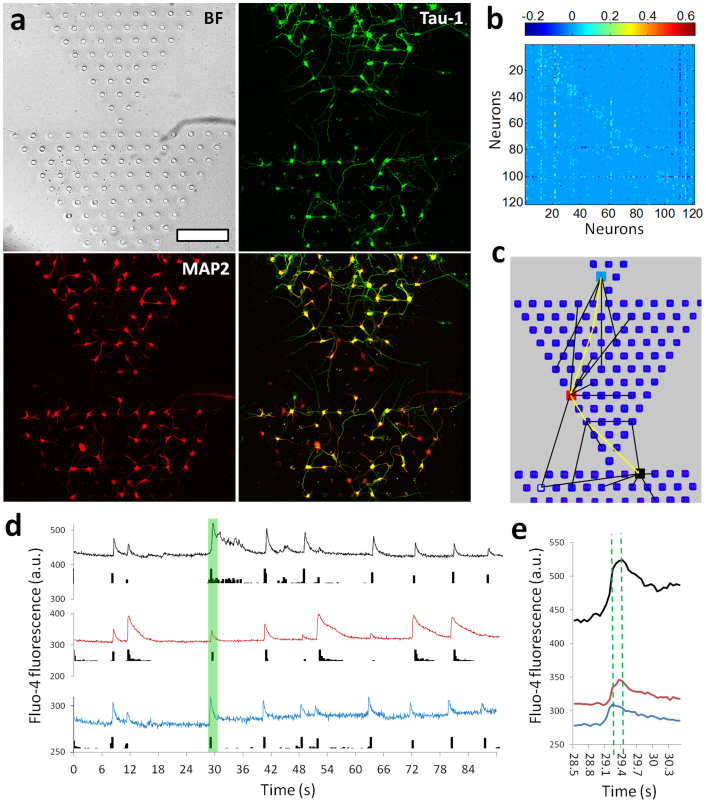
Figure 5. Patterned neural network assembly with a “diode” topology.
(a) Images showing the growth of neurons in a “diode” NeuroArray with series of triangle patterns. 5DIV neurons were fluorescent stained for Tau-1 (green) and MAP2 (red). Scale bar, 200 µm. (b) Connectivity matrix for a “diode” neural network with 120 functional units. (c) Illustrative diagram partially showing connections between different elements in a “diode” neural network. (d) Traces of calcium fluctuation of three neurons (color-coded in panel (c)) showing their spiking activities. (e) Details of fluorescent traces for the time window indicated by green box in panel (d).

