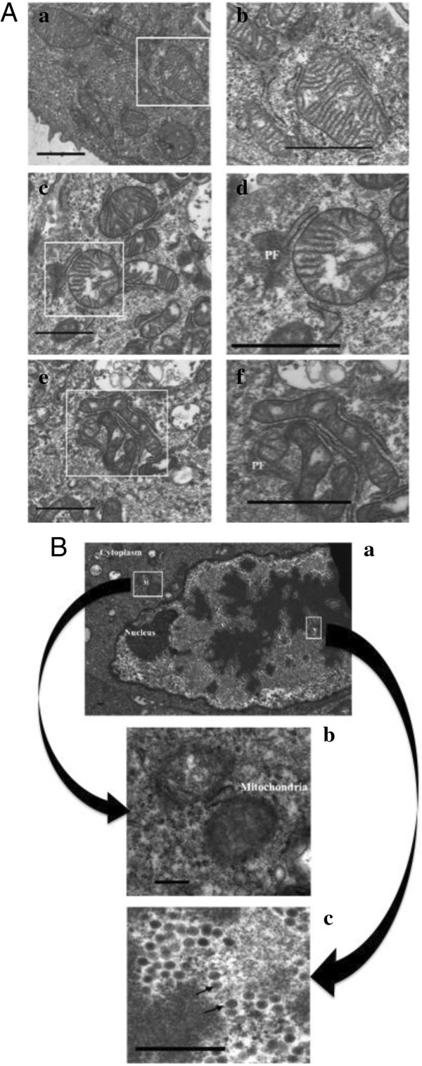
Figure 1.
Electron microscopy of BAdV-3 infected cells. (A) MDBK cells mock infected (panel a, b) or infected with wild-type BAdV-3 (panels c, d, e, f) at a MOI of 5 were analyzed after 6 (panel c, d) and 12 (panel e, f) hpi. Figures in the left panels (panels a, c, e) show the cytoplasm in the vicinity of the mitochondria. Area covered by white rectangles is enlarged and shown in the panels (panel b, d, f) on the right. Protein factories (PF) in the vicinity of the damaged mitochondria at 6 (panel d) and 12 (panel f) hpi. Bar = 0.25 μ. (B) MDBK cells infected with wild-type BAdV-3 at an MOI of 5 were analyzed at 24 hpi. An infected cell showing the virus particles (V) in the nucleus and the damaged mitochondria (M) in the cytoplasm (panel a). The enlarged area indicated by a rectangle (M) in panel “a” showing the mitochondria with amorphous internal structure in the cytoplasm of the infected cell (panel b). The enlarged area indicated by a rectangle (V) in panel “a” showing virus particles (indicated by arrows) in the nucleus of infected cells (panel c). Bar = 0.25 μ.

