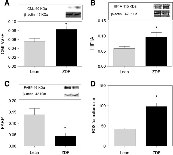
Figure 3.
Oxidative stress and damage in brains of ZDF rats. (A) Expression of CML/AGE is increased in ZDF compared to Lean. (B) HIF1A expression is higher in ZDF brain compared to Lean. (C) FABP is 3 times lower in ZDF rats compared to Lean. D) ROS formation is higher in ZDF brains compared to Lean. ROS formation is measured by the level of Fluorescin fluorescence. Western blot data showing protein of interest (upper part) and β-actin (lower part). Data are Means ± SEM (n ≥ 5 per group), * <0.0001 (difference to control gray bars), unpaired t-test. Western blot expression is normalized to β-actin; lanes of western blot insets are in the same order as in the X-axis.

