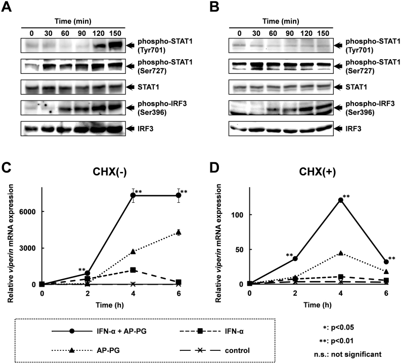
Figure 5. Phosphorylation of STAT1 at Ser 727 is immediately increased after stimulation with AP-PG.
(A), (B) RAW264.7 cells were pretreated with (A) or without (B) 5 mM cycloheximide (CHX) for 30 min. After the treatment, the cells were stimulated with 100 μg/ml of AP-PG. The cells were harvested at the indicated time points, and the whole cell lysates prepared from the cells were analyzed by immunoblotting using specific antibodies. Full length blots are presented in supplementary information (Supplementary Figure S1 and S2). (C), (D) RAW264.7 cells were pretreated with (D) or without (C) 5 mM cycloheximide (CHX) for 30 min, and then the cells were stimulated with 10,000 U/ml of IFN-α together with or without 100 μg/ml of AP-PG. After the indicated incubation periods, the cells were harvested, and the expression of viperin mRNA was analysed by real-time RT-PCR using a specific primer set. The data represent relative expression amounts compared with that at the initial time point after normalization with GAPDH mRNA expression. Double asterisk (**) show statistically significant differences (p < 0.01) compared to independent stimulations with IFN-α or AP-PG.

