
Figure 1.
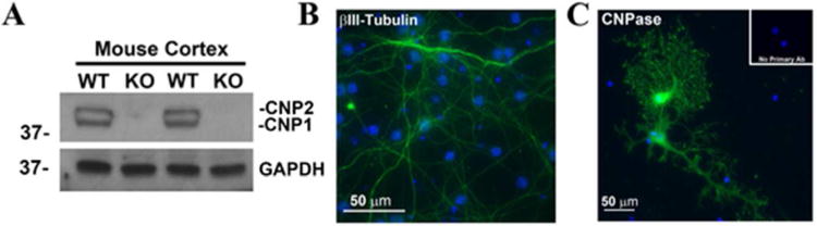
CNPase knockout confirmation and cell type identification. The complete lack of both type 1 (CNP1) and type 2 (CNP2) isoforms of CNPase was confirmed by Western blot on protein extracts from CNPase +/+ and -/- mouse cortical tissue using an anti-CNPase antibody. GAPDH is shown as a loading control (A). A neuron specific beta III-tubulin antibody and a FITC-conjugated secondary antibody was used to confirm pure cultures of primary mouse neurons (Green, DAPI, blue; Scale bar = 50 microns) (B). An anti-CNPase antibody was used to detect mature mouse oligodendrocyte cultures (Green, DAPI, blue; Scale bar = 50 microns) (C).