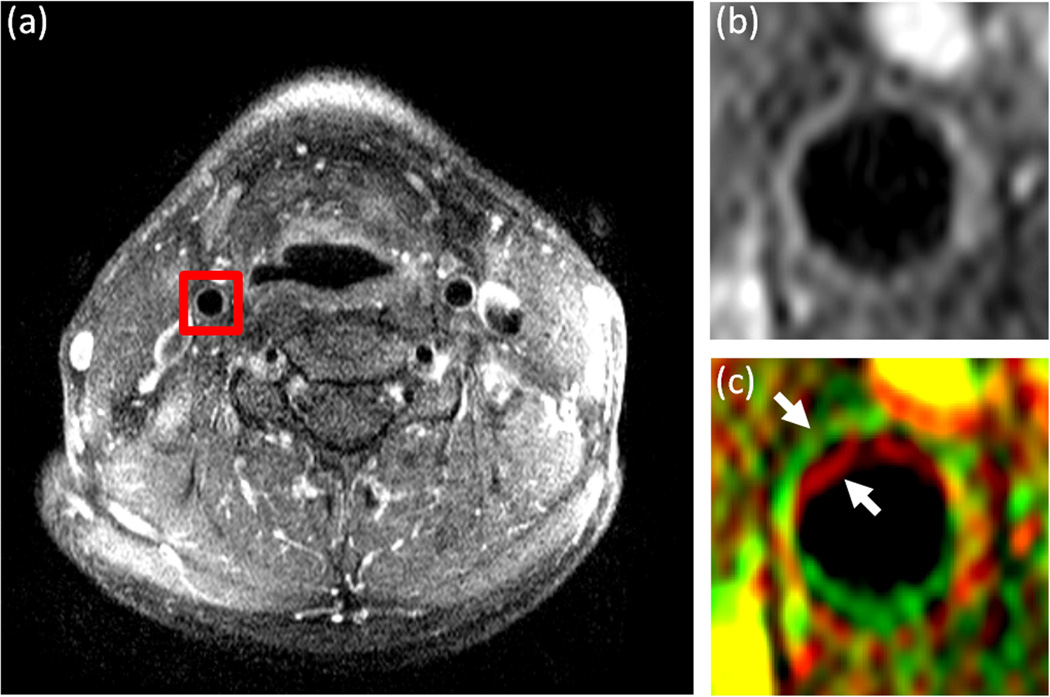
Figure 1.
DCE-MR images of the carotid arteries in the neck of a representative subject. a: A single image from a DCE-MRI series with a red square surrounding the right carotid artery indicating the cropped region used for analysis. b: The 14th frame in the DCE-MRI series used as the template image for registration and (c) the 15th frame in the series (rendered in green) overlaid on the 14th frame (rendered in red). The two consecutive frames are overlaid to show the misalignment between images in the series before registration. The arrows indicate the position of the carotid artery wall in each of the frames.

