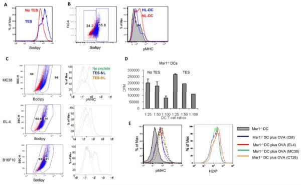
Figure 3. Effect of TES on antigen processing in DCs.
A. Typical example of lipid level in gated CD11c+ DCs treated with EL-4 TES for 48 h after staining with BODIPY. Seven experiments with similar results were performed. B. Cross presentation of antigens by DCs with different levels of lipids. DCs treated with TES and loaded with OVA were stained with BODIPY. Left panel - example of gates set for discriminating DCs with normal lipid levels (NL-DCs) from DCs with high lipid levels (HL-DCs). DCs were considered NL-DCs when their fluorescence overlapped the fluorescence of the control DCs. Control DCs in red; DCs treated with TES in blue. Three experiments, with the same results, were performed. C. Cross presentation of long OVA peptide by DCs with different levels of lipids. Left panels show the gate of NL-DCs and HL-DCs and pMHC expression is shown in the right panel. D. TES does not affect cross-presentation in CD204 deficient DCs. DCs generated from Msr1−/− mice were treated with TES, loaded with OVA, and used for stimulation of OT-1 T cells as described in Fig. 1C. Proliferation of OT-I T cells was measured in triplicate. Typical results of 4 performed experiments are shown. E. Typical examples of pMHC (left panel) and H2Kb (right panel) expression in CD204 deficient DCs. Grey shaded line - DCs without OVA treatment; blue line - DCs loaded with OVA in control medium (CM); red, green, and orange lines - DCs loaded with OVA and pre-treated with TES from EL-4, MC38, and CT26 tumors, respectively.

