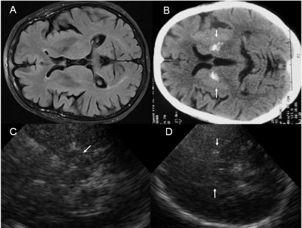
Figure 1.

Images of a patient with ET-PD-RLS, suggesting an alternative diagnosis of parkinsonism after paraclinical tests. A-cranial MRI, T2W, B-cranial CT, markedly hyperintensive signals bilaterally in the lentiform nucleus (white arrows), C-TCS, mesencephalic plane, moderate SN and red nucleus hyperechogenicity (white arrow), D-TCS, diencephalic plane, bilaterally markedly hyperechogenic lentiform nuclei (white arrows).
