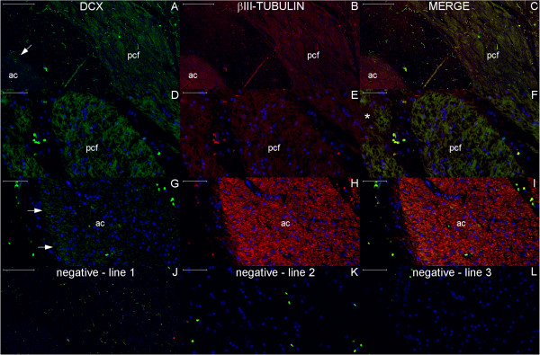
Figure 20.

DCX and βIII-tubulin expression in the post-commissural fornix and anterior commissure. For references for the locations studied in lines 1, 2 and 3, see Figure 19, numbers 6, 7 and 8, respectively. Figures (A–C) show that the post-commissural fornix (pcf) and the anterior commissure (ac) express both DCX (green) and βIII-tubulin (red). However, DCX expression is dense in the pcf (D); in the anterior commissure, DCX expression occurs only in the outer zone (arrows in (A) and (G)). Conversely, βIII-tubulin expression, which is sparse (asterisk in (F)) in the absence of DCX expression and weak in the pcf (E), is abundant in the anterior commissure (H). (J–L) are images of the negative controls in the counterpart locations. Scale bars: (A–C), (J) = 500 μm; (D–I), (K), (L) = 50 μm.
