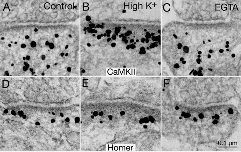
Fig. 10. CaMKII but not Homer is excluded from the PSD complex after EGTA treatment.
CaMKII label (top row) is typically dispersed in the postsynaptic compartment under basal control conditions (A), but aggregates at the PSD complex after high K+ depolarization (B; 90 mM for 2 min). Concentration of CaMKII label decreased in the PSD complex (area up to 120 nm from the postsynaptic membrane) under calcium-free conditions (C; 1 mM EGTA for 2 min). In contrast, the density and distribution of label for Homer 1 (bottom row) remain unchanged in control (D), high K+ (E) or EGTA (F). Scale bar =0.1 μm.

