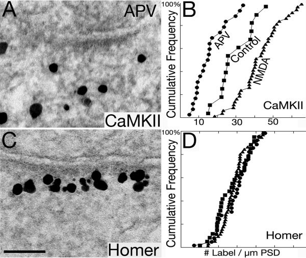
Fig. 11. CaMKII but not Homer is excluded from the PSD complex after APV treatment.
After APV treatment (50 mM, 2 min), the amount of CaMKII decreases, and sometimes it is absent from the PSD complex (A), while label for Homer 1 is consistently concentrated at the PSD (C). Cumulative frequency distributions (B, D) show significant differences in the density of label for CaMKII at PSDs between control, NMDA and APV treatment (B; P< 0.005 between control vs. NMDA, and control vs. APV, respectively). In contrast, the density of Homer 1 labeling does not differ significantly under the same experimental conditions (D).

