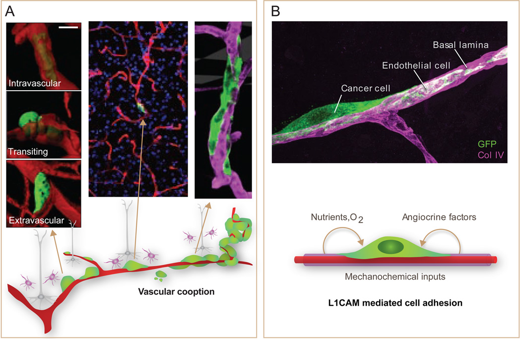
Figure 6. The perivascular niche for metastasis initiation.
A. Metastasis-initiating cells (green) exiting from brain capillaries (red) remain tightly associated with the vessels, adhering to and stretching over their abluminal surface. This interaction is required for metastatic outgrowth. Outgrowth occurs forming a furrow over the capillary and later a multilayered cell colony. B. The axonal guidance cell adhesion receptor L1CAM is aberrantly expressed in tumors, and its expression is associated with relapse. L1CAM in metastasis-initiating cells (green) that infiltrate the brain mediates their adhesion to capillary basal lamina (magenta). Besides providing MetSCs with mechanochemical cues, vascular cooption facilitates their access to oxygen and nutrients from the blood supply and to factors from the endothelium and surrounding stroma. [Adapted from (Valiente et al., 2014)].

