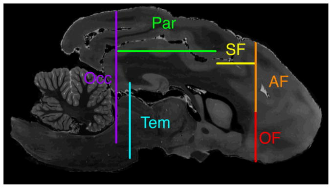
Figure 1. Relative ROI positions.

Sagittal T2-weighted MR image shows relative positions of cross sections of all ROIs. Orbital frontal ROI (OF) is shown in red, anterior frontal ROI (AF) in orange, superior frontal ROI (SF) in yellow, parietal ROI (Par) in green, occipital ROI (Occ) in purple, and temporal ROI (Tem) in blue.
