
Figure 2. Images showing use of histological sections to validate site for placement of anterior frontal (AF) and orbital frontal (OF) ROIs.
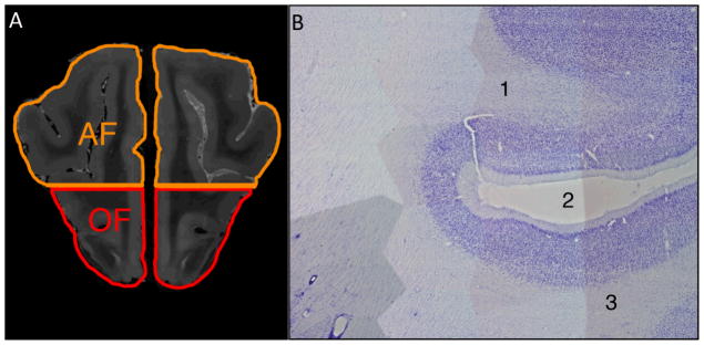
A) Coronal T2-weighted MR image located one-fourth of the distance from the genu to the anterior frontal cortex shows the AF and OF ROIs separated at the level of the inferior border of the pre-Sylvian sulcus, through which the horizontal line passes.
B) Parasagittal 50 micron Nissl-stained section of the canine brain that underwent MR imaging used to confirm ROI placement shows the characteristic features of the (1) AF region, (2) the pre-Sylvian sulcus, and (3) the OF region. Both the AF and OF typically have the prominent staining of layer II, poorly defined layer IV, and a thin layer V, with medium sized pyramidal neurons shown in this image. Identification of the pre-Sylvian sulcus and adjacent AF and OF regions confirmed proper placement of the ROIs shown in A.