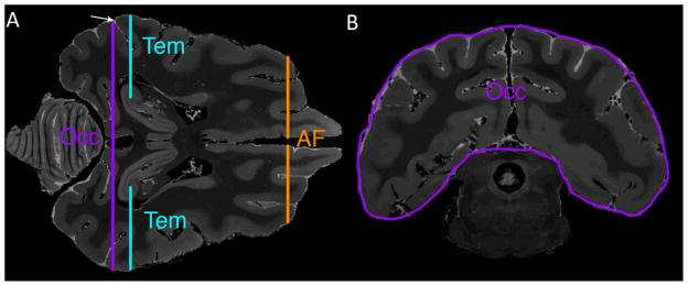
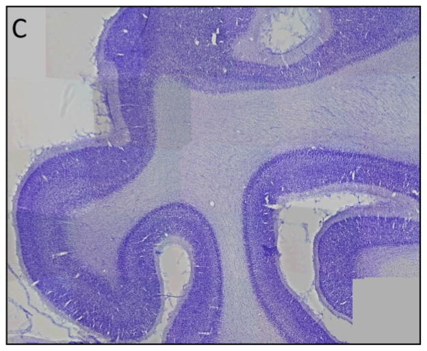
Figure 3. Use of histological sections to validate site for placement of occipital ROI.

A) Axial T2-weighted image at the level of the inferior splenium is used to identify the post-Sylvian sulcus marked by an arrow. The occipital lobe (Occ), temporal lobe (Tem), and anterior frontal (AF) ROIs are shown in cross section.
B) Coronal T2-weighted MR image at the level of the post-Sylvian sulcus shows the occipital lobe ROI (Occ).
C) Parasagittal 50 micron Nissl-stained section corresponding to MR images in A and B shows the characteristic appearance of the occipital cortex (i.e., prominent staining of layer IV), thus validating placement of the ROIs shown in A and B.

