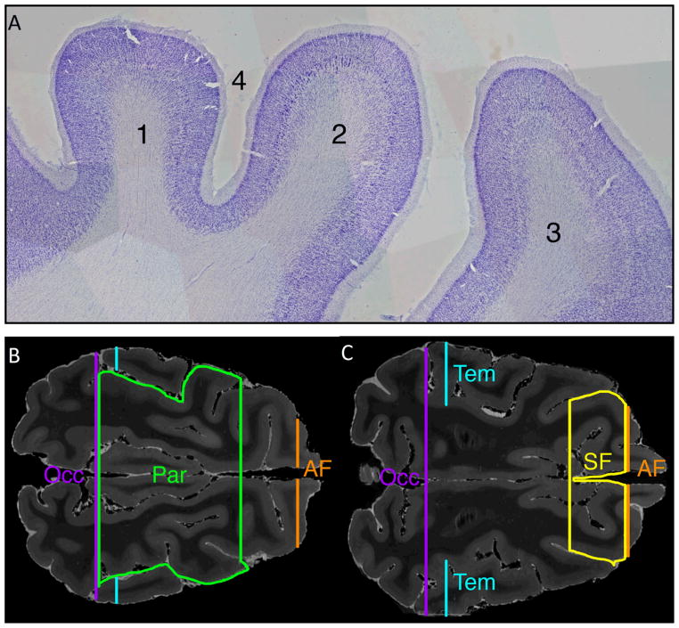
Figure 5. Images showing use of histological sections to validate site for placement of superior frontal (SF) and parietal lobe (Par) ROI placement.
A) Parasagittal 50 micron Nissl-stained section showing (1) parietal cortex with characteristic absence of large layer V pyramidal cells and Betz cells, (2) primary motor cortex (characterized by presence of large layer V pyramidal cells and Betz cells), (3) premotor cortex, and (4) post-cruciate sulcus. The sulcus separating the primary motor cortex and parietal cortex regions was used to delineate the SF and Par ROIs.
B) T2-weighted axial MR image at the level of the superior cingulate white matter shows the Par ROI in green. The histological features in A, demarcating the parietal cortex and frontal cortex, were used to confirm placement of the Par ROI, which is bounded posteriorly by the occipital (Occ) ROI, laterally by the supra-Sylvian sulcus, and anteriorly by the post-cruciate sulcus. Also shown in cross section are the Occ ROI (in purple), temporal ROI (Tem, in blue) and anterior frontal ROI (AF, in orange).
C) T2-weighted axial MR image at the superior border of the lateral ventricles shows the SF ROI in yellow. The SF ROI is bounded anteriorly by the AF ROI, laterally by the coronal sulcus, and posteriorly by the post-cruciate sulcus. Also shown in cross section are the Occ ROI (purple), Tem ROI (blue) and AF ROI (orange).

