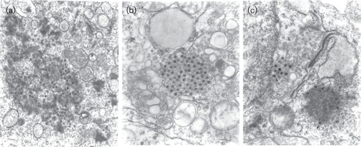
Fig. 1.

Electron micrographs of viruses SVIV (IAn-66411), TGV (JKT-8132) and TGV in BHK cells. Virions are shown by pointed arrows. (a) JKT-8132. Ultrastructure of reovirus fibrillar aggregate in the cytoplasm of a BHK cell with virus particles and cores. (b) IAn-66411 #5670. An aggregate of reovirus particles ~60 nm in diameter in the cytoplasm of a C6/36 cell, distance bar, 100 nm. (c) IAn-66411 #5672. A portion of a C6/36 cell infected with reovirus IAn-66411 showing viral protein aggregates with forming cores and virus particles ~60 nm in diameter (thick arrows) and microtubules (thin arrow) inside a cistern of granular endoplasmic reticulum which is expanded at one end. Bars, 100 nm.
