
Figure 1. Working mechanism of the reconfigurable plasmofluidic lens.
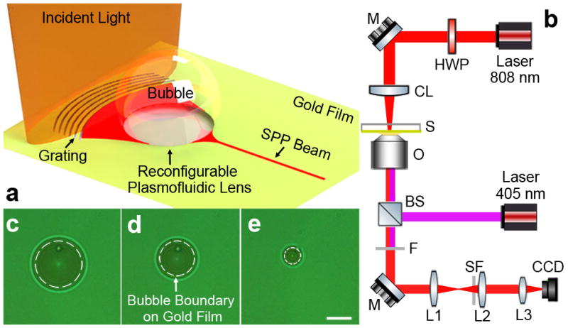
(a) Schematic of the reconfigurable plasmofluidic lens, wherein a laser-induced surface bubble is used to control the propagation of SPPs at the metal surface. (b) Schematic of the experimental setup. HWP: half wave plate; M: mirror; CL: cylindrical lens; S: sample; O: oil immersion objective; BS: beamsplitter; F: long pass filter; SF: spatial filter; and L: lens. The SPP field is imaged by leakage radiation microscopy which consists of a high-numerical-aperture oil immersion objective (NA=1.49), three lenses (L1, L2, and L3), and a charge-coupled device camera (CCD). A diode laser with a wavelength of 405 nm is coupled into the leakage radiation microscopy by a beamsplitter and focused on the gold surface through the same oil immersion objective. (c–e) Surface bubbles with different diameters (18, 14, and 6 μm, respectively) generated on the gold film. The white dashed circle represents the surface bubble boundary on gold film. Scale bar: 10 μm.