
Figure 3. Experimental demonstration of SPP divergence by the reconfigurable plasmofluidic lens.
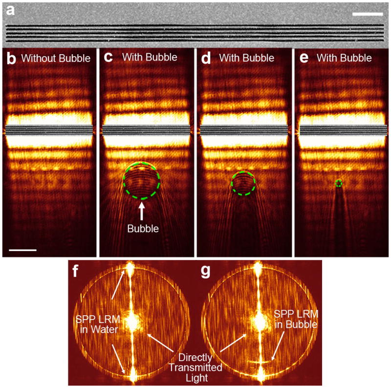
(a) Scanning electron microscope image of the fabricated grating. Scale bar: 4 μm. (b) Leakage radiation image of SPPs when surface bubbles are absent. The image was recorded at the image plane. Scale bar: 20 μm. (c–e) Leakage radiation image of SPPs propagating through three surface bubbles with different diameters (27, 17, and 5 μm, respectively). The green dashed circle represents the surface bubble boundary on gold film. (f–g) Fourier plane images of SPPs in the absence and presence of a surface bubble, respectively.