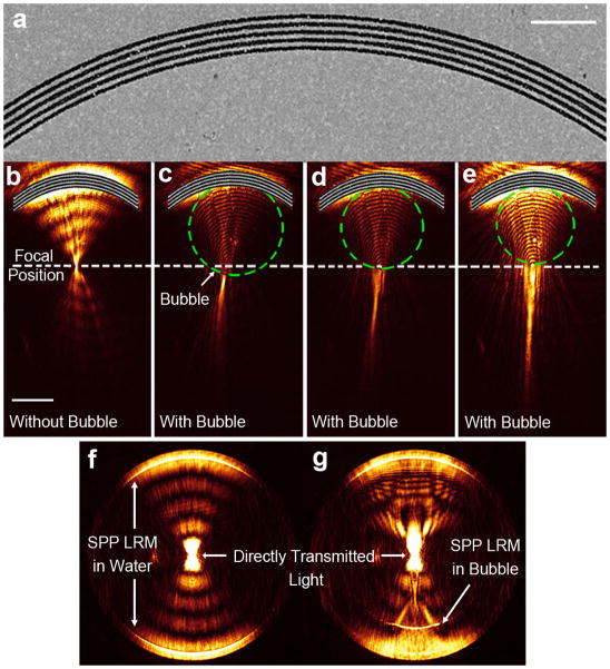
Figure 4. Experimental demonstration of SPP focusing and collimation by the reconfigurable plasmofluidic lens.
(a) Scanning electron microscope image of the fabricated arc grating. Scale bar: 5 μm. (b) Leakage radiation image of SPPs focused by the arc grating when surface bubbles are absent (the focal position is indicated by a white dashed line). (c–e) Leakage radiation image for SPPs propagating through three surface bubbles with different diameters (45, 39, and 37 μm, respectively). The image was recorded at the image plane. The green dashed circle represents the surface bubble boundary on gold film. Scale bar: 20 μm. (f–g) Fourier plane images of SPPs in the absence and presence of a surface bubble, respectively.

