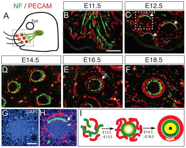
Figure 1. Nerve and blood vessels are organized into a “double ring” structure in the whisker follicle during development.
(A–F) Developmental profile of trigeminal axon innervation and blood vessel patterning in the whisker follicles. Trigeminal axons and blood vessels are visualized by neurofilament (green) and PECAM (red), respectively, in the snout area (boxed region in Fig. 1A). Co-immunostained tangential sections of whisker follicles at E11.5 (B), E12.5 (C), E14.5 (D), E16.5 (E), E18.5 (F) show the dynamic processes that lead to the stereotypic organization of a double ring structure. The trigeminal axons form a ring-like structure as early as E12.5 (arrowheads in C), and are then surrounded by a blood vessel ring that results in the formation of a double ring structure around each follicle at E16.5, with the nerve ring located inside and the vessel ring outside (arrow in E). (G–H) One whisker follicle in C (dotted box) is overlaid by DAPI staining to show follicle primordium (dotted circle in G). Nerve terminals surround the outside of the primordium (arrow in H). (I) Schematic illustration of nerve and vessel ring organization in the whisker follicle. Scale bar: 100 μm (B–F), 50μm (G–H). See also Figure S1 and Movie S1–S3.

