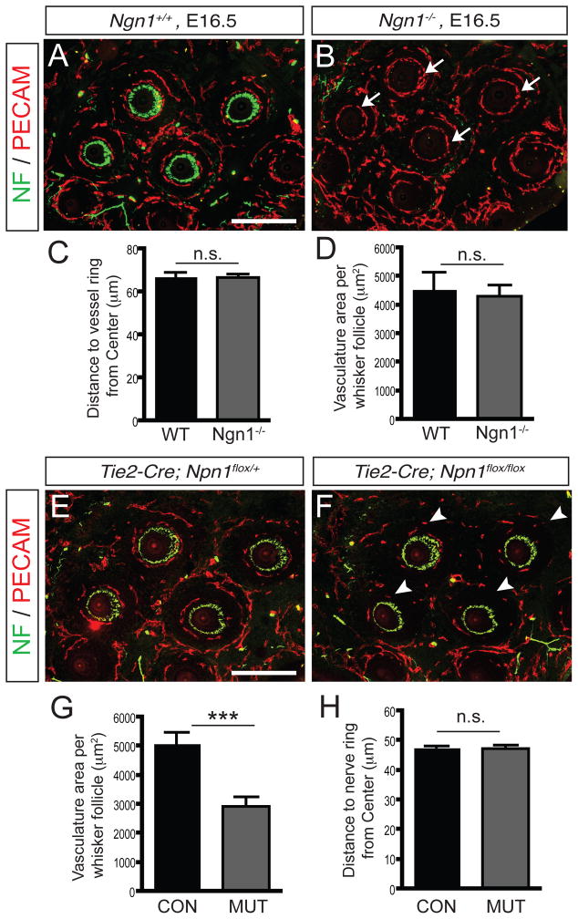
Figure 2. Nerve ring and vessel ring are patterned independent of each other in the whisker follicle.
(A–B) Nerve/vessel immunostaining of wild-type littermate control exhibits normal double ring patterning (A). Nerve/vessel immunostaining of the Ngn1 knockout shows no trigeminal innervation in the whisker follicles, but blood vessel rings are normally organized at each whisker follicle (arrows in B, C–D). (C–D) For the quantification, 10 pairs of whisker follicles were analyzed and the mean distance from the center (C) and vasculature area surrounding the follicle (D) ± SEM is shown. (E–F) Endothelial specific-Npn1 deletion shows less vasculature formation around whisker follicle (white arrowheads in F and G) compared to the vasculature of control (E), but nerve rings are organized normally (H). (G–H) Vasculature area surrounding follicle (G) and mean distance from center to nerve ring (H) ± SEM is shown (n=14). Paired student t-test; ***, P<0.001; n.s, not significant. Scale bar: 200 μm (A–B and E–F)

