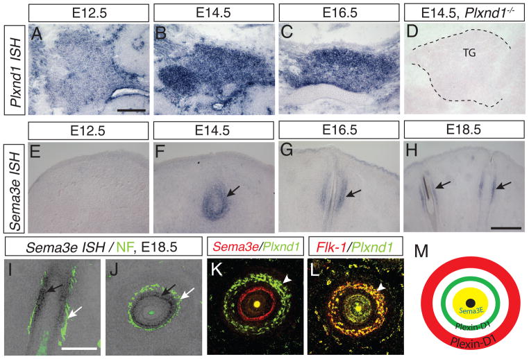
Figure 3. Complementary expression pattern of Plexin-D1 in the TG and blood vessels and Sema3E in the whisker follicle.
(A–D) Plxnd1 in situ hybridization (ISH) on sagittal sections of wild-type embryos at E12.5 (A), E14.5 (B), and E16.5 (C) shows that Plxnd1 mRNA is detected at very low level as early as E12.5 and is significantly increased at E14.5 and continues to E16.5 as the double ring structure develops. Plxnd1 mRNA is not detectable in sections of the trigeminal ganglion from plexin-D1 knockout animals at E14.5 (TG outlined by dashed line in D). (E–H) Sema3e mRNA ISH on coronal sections parallel to the whisker follicles at E12.5 (E), E14.5 (F), E16.5 (G) and E18.5 (H) shows that Sema3e mRNA is expressed in the mesenchymal sheath surrounding the hair follicles starting at E14.5 and continues at E16.5 and E18.5 (black arrows). Scale bar: 100 μm in A applies to G, and 200 μm (H) (I–J) Neurofilament immunostaining (white arrows) after Sema3e ISH on the same sections shows that Sema3e (black arrows) is expressed inside of the nerve rings (coronal image in I and tangential image in J). (K–L) Double fluorescence ISH with Plxnd1 and Sema3e (K) or Flk-1 (L) shows that Plxnd1 mRNA is expressed in the endothelial cells that form the vessel ring (white arrowhead in K and L) and Sema3e is expressed inside of both nerve and vessel rings. (M) Summary of Plxnd1 and Sema3e mRNA expression pattern in the whisker follicle. Sema3e is expressed in mesenchymal tissue (yellow) and Plxnd1 is expressed in blood vessel (red) and in TG therefore Plexin-D1 protein probably in nerve (green). Scale bar: 100 μm in A applies to G, 200 μm (H), and 100 μm in I applies to L.

