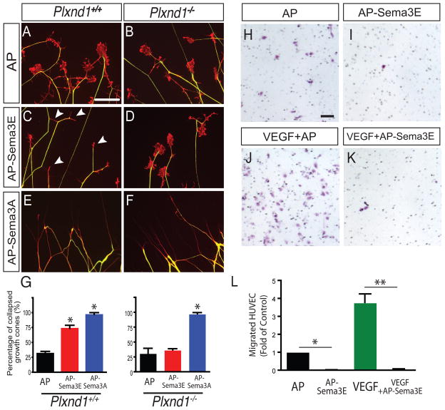
Figure 4. Sema3E-Plexin-D1 signaling provides a repulsive guidance cue to both TG axons and blood vessels in vitro.
(A–G) Sema3E-Plexin-D1 signaling serves as repulsive cue for cultured TG neurons. Trigeminal ganglia were isolated from wild type (A, C, E) or Plexin-D1 null mice (B, D, F) at E14.5 and a growth cone collapse assay was performed. Axons (green) and growth cones (red) were visualized by anti-neurofilament and phalloidin staining, respectively. After incubation with 2 nM of AP (A, B), AP-Sema3E (C, D), or AP-Sema3A (E, F) for 30 min, >200 growth cones were scored for each experimental condition. Sema3E treatment induced significant growth cone collapse (white arrowheads in C), but growth cone collapse was absent in Plexin-D1 mutant TG neurons (D). (G) Quantification of growth cone collapse assay (n=3, data shown as mean ± SD, ANOVA; *, P<0.001). (H–L) Sema3E inhibits endothelial cell migration. VEGF-induced HUVEC transwell migration assay was performed in the presence (K) or absence (J) of Sema3E (AP-Sema3E, 0.5 nM). After a 5 hr incubation, migrated cells were fixed and visualized by Nissl staining. Sema3E prevents HUVEC migration induced by VEGF as well as the basal level of migration (I). (L) Quantification of migrated HUVECs (data shown as mean ± SEM, n=3, ANOVA; *, P<0.05; **, P<0.001). Scale bar: 50 μm (A–F, H–K). See also Figure S2.

