Figure 1.
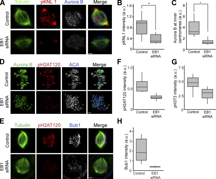
EB1 localizes Aurora B to centromeres to phosphorylate kinetochores. (A) HeLa cells depleted of EB1 were immunostained with antiphospho-KNL1(Ser60) (pKNL1) and Aurora B antibodies. Bar, 2 µm. (B) Quantification of immunostaining intensities of pKNL1 shown in A. *, P = 2.0 × 10−127. (C) Centromeric Aurora B levels measured in control and EB1-depleted cells (n > 300 centromeres). *, P = 1.2 × 10−105. (D) EB1 depletion reduces Bub1-mediated phosphorylation of histone H2A (pH2AT120). Bar, 1.6 µm. (E) EB1 depletion reduces Bub1 at kinetochores. Bar, 2.2 µm. (F–H) Quantification of pH2AT120 (F), phospho–histone H3Thr3 (pH3T3; G), and Bub1 (H) levels in control and EB1-depleted HeLa cells (see Fig. S1 B for pH3T3 staining examples). The height of the boxes represents the interquartile range (IQR). The central horizontal lines depict the median. The top whiskers represent the 75th percentile + 1.5× IQR, and the bottom whiskers represent the 25th percentile − 1.5× IQR. ACA, anticentromere antigen; a.u., arbitrary unit.

