Figure 7.
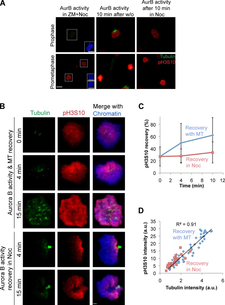
Aurora B activity on chromatin is regulated by microtubules in prometaphase but not in prophase. (A) Phospho–histone H3Ser10 (pH3S10) and tubulin immunostaining of prophase and prometaphase cells at the indicated conditions. Insets show chromatin staining. AurB, Aurora B; w/o, washout. (Assay scheme is shown in Fig. S5 B.) Bars, 10 µm. (B) HeLa cells in prometaphase immunostained for tubulin, phospho–histone H3Ser10 (pH3S10), and chromatin at the indicated conditions. Bar, 2.2 µm. (C) Relative pH3S10 intensity plotted as a function of time after ZM washout in the presence or absence of nocodazole. Error bars show standard deviations. (D) Correlation between total cellular pH3S10 intensity from all time points and total tubulin intensity measured per cell (R2 = 0.91). Cellular pH3S10 intensities at 0 min and recovery phases in the absence of nocodazole (Noc) are represented as blue diamonds, and intensities from cells in nocodazole are shown in red squares (n = 58; data shown are one representative experiment of three repeats). MT, microtubule; a.u., arbitrary units.

