Figure 4.
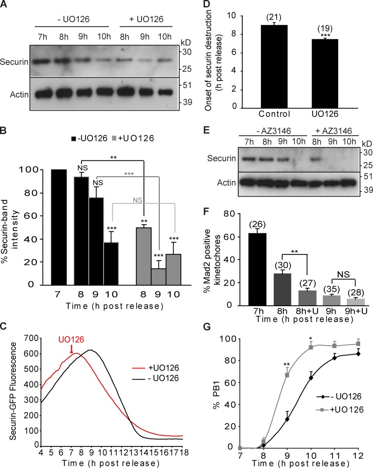
Inhibition of MAPK in late prometaphase I induces early destruction of securin, faster Mad2 dissociation, and accelerated PB1 extrusion. (A and B) Western blot (20 oocytes/lane; A) and analysis of securin (B) in oocytes at 7, 8, 9 and 10 h after release from IBMX, in the presence or absence of UO126. (C) Representative fluorescence traces of oocytes injected with securin-GFP in the presence or absence of UO126. (D) Time of onset of securin-GFP destruction in the UO126-treated oocytes (n = 19 from two experiments) and nontreated control oocytes (n = 21 from two experiments). The peak of the graph was taken as a marker for the start of destruction. (E) Western blot (20 oocytes/lane, n = 2) of securin in oocytes at 7, 8, 9, and 10 h after release from IBMX, in the presence or absence of AZ3146. (F) The proportion of Mad2-positive kinetochores at 7, 8, and 9 h after release from IBMX, in the presence or absence of UO126 (U). (G) Timing of PB1 extrusion in control (n = 116) and UO126-treated oocytes (n = 112). In B, D, F, and G, results were compared with the nontreated controls or to each other where indicated and expressed as SEMs. UO126 and AZ3146 were added to the media at 7 h after release from IBMX. NS, P > 0.05; *, P < 0.05; **, P < 0.01; ***, P < 0.001.

