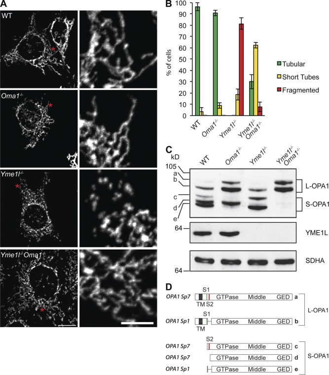
Figure 1.
Loss of OMA1 restores tubular mitochondria in Yme1l−/− cells. (A) Representative images of mitochondrial morphology in MEFs. The asterisks denote the area of magnification enlarged on the right. Bars: (left) 15 µm; (right) 5 µm. (B) Quantification of three independent experiments (error bars indicate mean ± SD), n ≥ 100. (C) Accumulation of OPA1 forms in MEFs lacking YME1L, OMA1, or both. Cells lysates were analyzed by SDS-PAGE analysis and immunoblotting using the indicated antibodies. a–e, OPA1 forms. (D) Schematic representation of mature L-OPA1 forms derived from splice variants 1 and 7 and S-OPA1 forms produced by cleavage at proteolytic sites S1 or S2 by OMA1 or YME1L, respectively.

