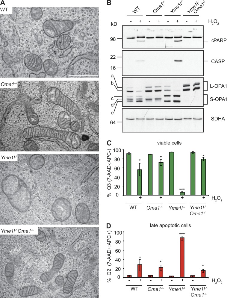
Figure 3.
Loss of OMA1 restores cristae morphology and apoptotic resistance of Yme1l−/− cells. (A) WT, Oma1−/−, Yme1l−/−, and Yme1l−/−Oma1−/− MEFs were analyzed by transmission electron microscopy. Bar, 1 µm. (B–D) WT and protease-deficient MEFs were exposed to H2O2 to induce apoptosis. (B) Immunoblot analysis of apoptotic marker proteins (cleaved PARP, cPARP; caspase 3, CASP). (C and D) Flow cytometry analysis of viable cells (Q3: 7-AAD−;APC−) and late apoptotic cells (Q2: 7-AAD+; APC+). *, P ≤ 0.05; ***, P ≤ 0.001. Error bars indicate mean ± SD.

