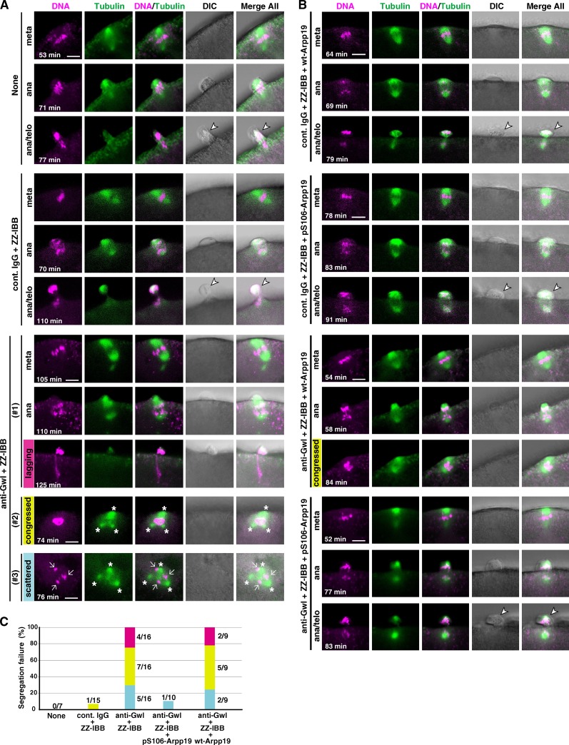
Figure 4.
Chromosome segregation in meiosis I is abortive in Gwl-inhibited oocytes. (A and B) Immature starfish oocytes were first injected with HiLyte 488–labeled tubulin (green) and Alexa 568–labeled histone H1 (magenta), and then injected with neutralizing anti-Gwl antibody (anti-Gwl) or control IgG (cont. IgG) along with ZZ-IBB (that can deliver cytoplasmically injected IgG into the nucleus) or uninjected (None). After 1-MeAde addition, live-cell images were obtained using a confocal microscope to monitor formation of the meiosis I spindle. Gwl-inhibited oocytes failed to properly segregate homologous chromosomes (A). This phenotype may be classified into three types, although they overlapped in some cases: lagging (#1, magenta), congressed (#2, yellow), or scattered (#3, blue) chromosomes along with frequently multipolar spindles (asterisks). However, further coinjection into immature oocytes with Arpp19 that had been in vitro thiophosphorylated by Gwl (pS106-Arpp19), but not with wild-type Arpp19 (wt-Arpp19), restored homologous chromosome segregation (B). Time after 1-MeAde addition is indicated in each frame. Arrow, chromosomes; arrowhead, the first polar body. Bars, 5 µm. (C) Quantification of chromosome segregation failure displayed in A and B. Each color corresponds to #1, #2, and #3 in A and B, respectively. Numbers of independent experiments (more than three females) are indicated at each bar.

