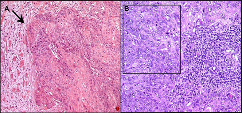
Figure 1.

(A) Histopathological analysis of cervical tissue. Nests of neoplastic squamous cells are invading the stroma (arrow head). The cancer is poorly differentiated and keratinizing (original magnification ×200). (B) Pelvic lymph node. Metastatic cells from squamous cell carcinoma (black square) are surrounded by normal lymphocytes (original magnification ×200).
