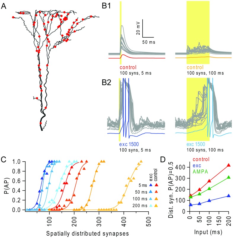
Figure 4. Background excitatory input enables integration of spatially distributed synaptic input over extended temporal windows.
(A) Apical tuft with an example distribution of 60 synaptic inputs (red dots). (B1) Left: example voltage traces from all terminal apical branches (grey traces, N = 28) and the soma (red) in response to stimulation of 100 spatially distributed synaptic inputs at 200 Hz for 5 ms, during control conditions. Right: example trial for 100 ms stimulation window. (B2) Same as B1 in the presence of background activity from 1500 excitatory synapses. Somatic voltage traces are in blue and are truncated at +10 mV. (C) Probability of triggering action potentials (P(AP)) versus number of stimulated synapses, spatially distributed over the apical tuft. Synapses were driven with random trains with a window of 5 ms, 50 ms, 100 ms and 200 ms, and the frequency was scaled to maintain ∼1 quantal conductance per synapse, in the absence (red-yellow traces) and presence of background excitatory input (blue traces). (D) Number of distributed synapses required to trigger an AP with 50% probability (P(AP) = 0.5), for different levels of temporal dispersion in the stimulus evoked synapses (input window), in the absence (red) and presence of background excitatory input (blue), and during AMPAR-only background synaptic inputs which depolarized the dendrites to comparable levels (green).

