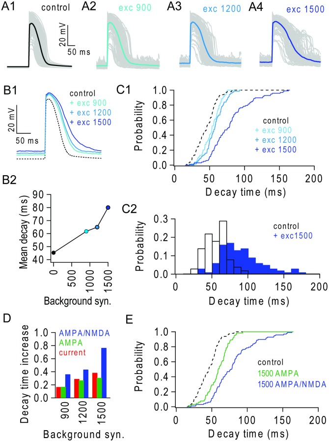
Figure 6. Background excitatory input extends the duration of NMDAR spikes.
(A1–4) Dendritic NMDAR spikes triggered in different terminal branches (30 synapses; 100 trials) in the absence (control) and presence of 900, 1200 and 1500 background excitatory synapses. Single trials (grey) and average (solid colour). (B1) Average NMDAR spikes in (A) overlaid. (B2) Average decay time (37% of peak) of NMDAR spikes in (A). (C1) Cumulative distributions of decay times for control and different levels of background input. (C2) NMDAR spike decay time distribution in the absence (black) and presence (blue) of 1500 background synapses. (D) Fractional increase in average NMDAR spike decay time for excitatory background synapses containing AMPAR/NMDARs (blue) and equivalent dendritic depolarization obtained with background AMPAR-only synapses (green) or current injection (red). (E) Cumulative distributions of NMDAR spike decay times during depolarization mediated by AMPAR-only (green) and mixed AMPAR/NMDAR (blue) background synaptic input.

