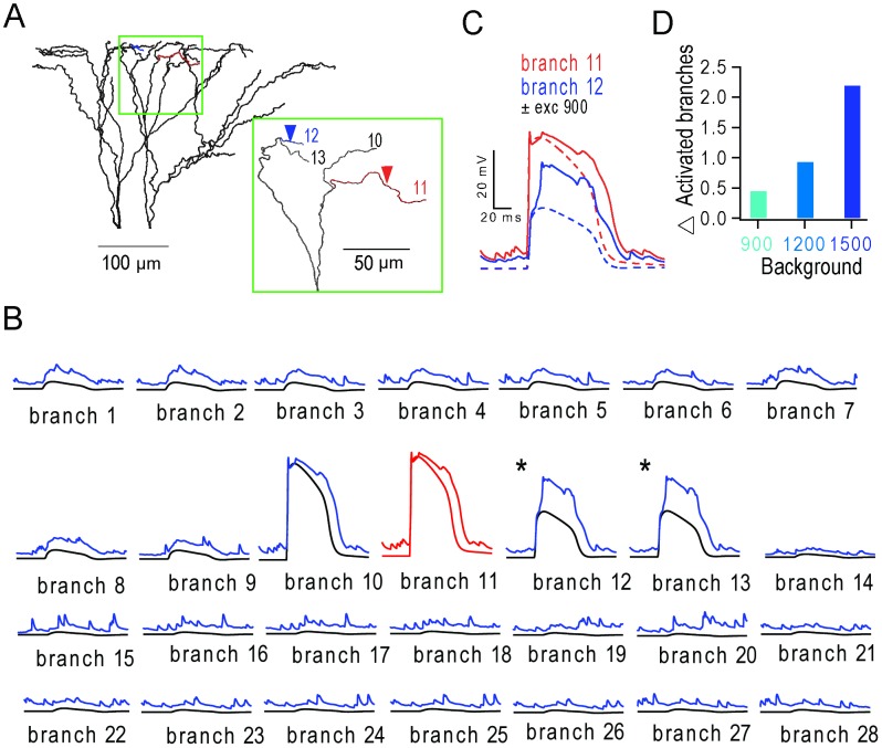
Figure 7. Stimulation of a dendritic branch triggers regenerative potentials in neighbouring branches during background synaptic input.
(A) Apical tuft with inset showing branch 11 (red) stimulated with 30 synaptic inputs (5 ms window) and branch 12 (blue) that receives no stimulus evoked input. Inset: enlarged tuft region with overlapping branches removed for clarity. (B) Membrane voltage of all 28 terminal apical branches during activation of branch 11 (red trace) in the presence (upper trace) and absence (lower trace) of distributed background synaptic activity from 900 excitatory inputs. Asterisks denote additional regenerative events triggered in branches 12 and 13 in the presence of background activity. (C) Voltage in branch 11 (red) and branch 12 (blue) with (solid lines) and without (dashed lines) background activity. (D) Average number of additional regenerative events triggered in neighbouring branches (identified using a 13 mV increase above level observed in the absence of background input) during different levels of background excitatory input (10 trials per branch, per condition).

