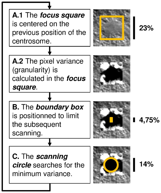
Figure 2. Tracking engine of the ACT macro.

A. The position of the centrosome in the previous frame is projected on the current frame. A square window is drawn around the projected position, and the granularity (i.e., pixel-value variance within a circle of about 1% of the embryo height) is determined within this window. The macro defines the size of this focus square to be large enough for the subsequent scanning of a circular region with minimal granularity. B. To make this scanning process efficient (and also to avoid too strong movements of the centrosomes from one frame to another), a smaller rectangular box is defined that limits the search within the boundaries of the box. We refer to this box as the boundary box. The height (hbb) is about 4% of the height of the embryo. (The width of the boundary box is half of its height.) The boundary box is centered vertically on the previous centrosome position. Horizontally it is shifted either towards the anterior or posterior side of the embryo to take into account the natural dynamic behavior of centrosomes; see Table S2 for more details. C. The actual search is done by positioning the center of a circle (which diameter, dsc, is about 14% of the height of the embryo) successively to all pixels inside the boundary box. At each pixel, the mean granularity within the scanning circle is determined. The pixel for which the circle surface area shows the minimal mean granularity marks the new center of the centrosome. This position is projected to the next frame, and the tracking engine continues with step A).
