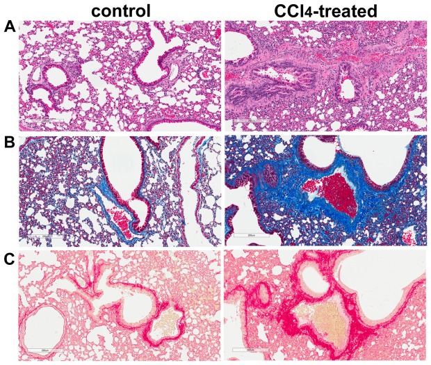
Figure 6. Marked collagen deposition is observed in lungs of the CCl4-treated mice.
A. H&E-stained lung sections. Representative bright-field micrographs of H&E-stained lung sections from control and CCl4-treated mice. B. Trichrome-stained lung sections. Representative images of trichrome-stained lung sections identify increased extracellular matrix deposition in lungs of CCl4-treated mice. C. PSR-stained lung sections. Representative bright-field micrographs of PSR-stained lung sections illustrating increased collagen deposition in lungs of CCl4-treated mice. Scale bar = 200 µm.

