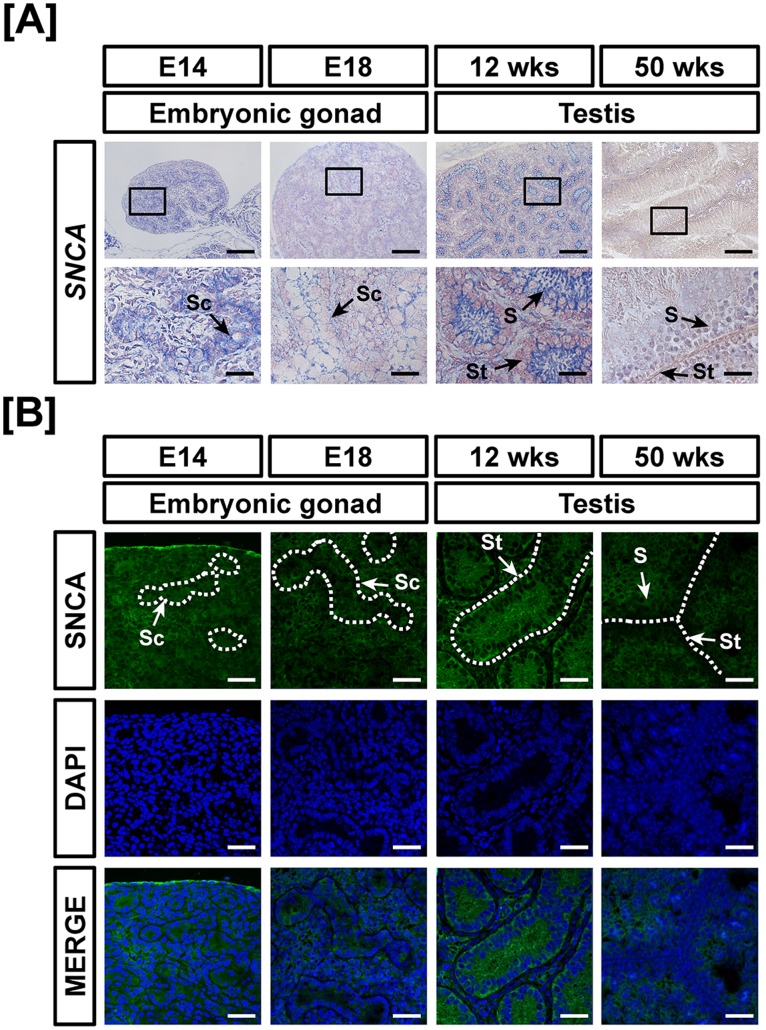
Figure 3. Cell-specific localization of mRNA and protein for SNCA in male reproductive tracts during their development.
Localization of SNCA expression was analyzed in the male reproductive tract of chickens during their development by in situ hybridization (A) and immunofluorescence analyses (B). Cell nuclei were stained with DAPI (blue). Legend: S, Sertoli cell; Sc, seminiferous cord; St, seminiferous tubule. Scale bar represents 100 µm and 20 µm for first and second horizontal panels of (A) and 50 µm for (B). See Materials and Methods for a complete description of the methods.

